The first trimester extends to the 14th week of pregnancy. Here are the main stages of embryonic and fetal development.
The first trimester extends to the 14th week of pregnancy. The embryo forms. Your baby’s first organs appear and their heart begins to beat. They’re only 9 cm long, but their head, torso, arms, and legs are clearly visible at the end of the first trimester. Here are the main stages of development, week by week.
Consult our fact sheets on fetal development during the second and third trimesters for more information.
This fact sheet focuses on the baby’s development. To learn more about how mothers experience the first trimester, consult our fact sheet entitled First trimester: Physical changes and common discomforts.
Week 1: Why 40 weeks of pregnancy?
The exact day that fertilization occurs is difficult to pinpoint. To calculate how far along you are, doctors go by the number of weeks without menstruation. Your due date is therefore counted from the first day of your last period. Consequently, 14 days are added to the length of your pregnancy. According to this method, you aren’t actually pregnant during the first 2 weeks.
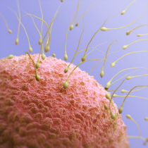
Week 2: Ovulation
Ovulation is the moment when one of your ovaries (left or right) expels an egg. Women typically ovulate 14 days before the first day of their next period. The expelled egg descends along the fallopian tube, which is the site of fertilization. It then continues to the uterus.
An egg’s lifespan is 12–24 hours. Your fertile period lasts about 5 days every month, from 4 days before ovulation to 24 hours after. Sperm can live for 3–5 days in a woman’s body.
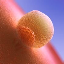
Week 3: Fertilization
A sperm enters the egg: they fuse to become an egg composed of a single cell. This is the moment of
fertilization and the true beginning of pregnancy.
The egg will take several months to develop into a baby. But it already contains all the necessary genetic information: sex, eye colour, height, skin pigmentation, etc. All this in a sphere the size of a pinhead!
During its first days of life, the egg descends slowly from the fallopian tube to the uterus. During this journey, the egg will divide into two cells. These will then divide to produce two more. These four cells will then become eight, and so on. This is the gradual process by which all of a human being’s organs, limbs, and systems are formed.
In rare cases, the egg will implant outside the uterus, most often in a fallopian tube. This is called an ectopic or tubal pregnancy. When this occurs, the pregnancy must be terminated to protect the health of the mother.
Twins
Identical twins come from a single fertilized egg that has completely separated into two embryos. They share the same genetic makeup, which means they will bear a striking resemblance to one another and necessarily be of the same sex. Fraternal (non-identical) twins, on the other hand, are formed by the fertilization of two eggs. They may therefore be of different sexes.
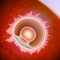
Week 4: Egg implantation
The fertilized egg attaches to the wall of your uterus. This stage is called implantation. It lasts a few days and requires the production of a special hormone produced by the egg: the hCG hormone. This is what a pregnancy test detects. Your hCG levels will double every 2–3 days in early pregnancy, peaking at around 8–10 weeks.
The presence of hCG leads to significant production of another hormone, progesterone. Its role is to keep the thickened lining of the uterus in place. The egg burrows deeper and deeper into the uterine wall to derive nutrients and oxygen from its many blood vessels.
At this stage, the egg has a diameter of 4–5 mm. It contains a number of cells and resembles a raspberry. It is made of two layers: the inner part will form the embryo, while the outer part will become the placenta, the envelope that protects and nourishes the embryo. Already, the first stage of the nervous system is in place.
Week 5: The umbilical cord
During week 5, the embryo’s round shape becomes elongated. It now looks like a tiny seahorse, measuring about 2.5 mm from crown to rump. The embryo grows quite a bit during this period, due in part to its developing circulatory system. It is now visible to the naked eye. The heart has entered its first stage of development. Little by little, your baby’s face takes shape: the eyes, nose, mouth, and ears.
The cells then organize themselves into three groups. Each will develop into different parts of your baby’s body:
-
Skin, eyes, ears, and nervous system
-
Muscles, skeleton, and blood vessels
-
Digestive tract, pancreas, and lungs
The umbilical cord now connects you to the embryo. It allows the embryo to draw nutrients from your diet as well as oxygen. The cord also allows for the evacuation of waste. Nestled in the uterine cavity, the embryo is surrounded by amniotic fluid.
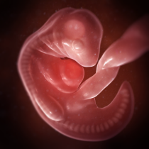
Week 6: Baby’s heart begins to beat!
During this week, 4 weeks after fertilization, an important part of the embryo will have developed: the
neural tube. Your baby’s entire nervous system will develop from this structure—their brain, at one end of the tube, as well as their spinal cord and nerves. By the end of this week, the ends of the tube will close. One of the purposes of taking folic acid supplements before and during pregnancy is to prevent neural tube defects, such as
spina bifida.
Week 6 is also when baby’s heart begins to beat. This can be observed via ultrasound. But that tiny heart is still forming. For now, it looks like a small bump, one that’s very clearly visible at the front of the embryo.
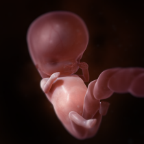
The embryo resembles a bean, with small arm and leg buds and the beginnings of a neck. It measures about 5 mm from crown to rump. The eye lenses and the inner ear begin to develop, as do the lungs, stomach, liver, and pancreas.
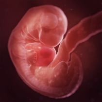
Week 7: All about the brain
This week is marked by significant brain development. The cortex begins to take shape with the development of the two hemispheres, the cerebral cortex, and grey matter.
Your baby’s head is forming gradually. The back of the brain becomes wider, causing the head to tilt forward.
It’s now possible to distinguish nostrils. The gums, where baby teeth will soon emerge, have already formed. There’s a dark area on either side of the head. Those are your baby’s eyes! Retinal pigment is what makes them a darker colour.
The vital organs are all in their earliest stages, though not yet functional. It’s possible to distinguish the four cavities of your baby’s heart. Your baby-to-be now measures about 10 mm from crown to rump.
Week 8: Movement begins
Your baby’s eyelids and outer ears (which look like small dimples) begin developing. However, the first senses to “awaken” will be smell and taste. At this stage, baby’s nose and upper lip have already formed. Week 8 is when receptors that can detect odours begin to appear.
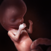
Little buds at the end of the limbs will develop into fingers and toes. It’s also possible to make out elbows and knees. No longer static, your baby now starts to move their torso and limbs. You won’t be able to feel it, but they’re moving within your uterus.
Several vital organs—the stomach, intestines, pancreas, and kidneys—start to take shape. At this point, they’re still not functional.
At the end of this week, your baby measures about 12 mm.
Week 9: Fingers and toes are now visible
During this week, your baby’s arms and legs become longer and their elbows and knees, along with their wrists and ankles, become more defined. Their muscles and nerves are also taking shape.
A baby’s head is always large compared to the rest of their body. But it gradually grows rounder, and the neck grows straighter. There are two dark zones which will become your baby’s eyes (for the moment, they’re widely spaced at either side of the head), two dimples for their ears, and only one opening for their nose and mouth.
On the sensory level, there are now touch receptors around the mouth. Your baby’s first taste buds will also develop during this week.
Depending on the sex of your child, the ovaries or testicles will begin to form. However, the genital organs won’t appear until the third month of pregnancy.
The embryo is looking more and more like the baby you’ll welcome into the world. The limbs continue to grow, with all their parts, including the joints, becoming clearly visible. The toes and fingers are well defined. Baby can now carry out a whole range of movements! They measure about 17 mm from crown to rump.
Week 10: A rapid heartbeat
Your baby’s eyelids have now formed, and they protect their developing eyes. The outer ears, meanwhile, are now very similar to yours! However, they sit slightly lower, close to the neck. Your little one measures about 35 mm from crown to rump and weighs around 8 g.
At this stage of pregnancy, your baby’s heart rate is around 170 beats per minute. It will slowly decrease as the months go by. It’s possible to hear your baby’s heartbeat using a device called a fetal doppler.
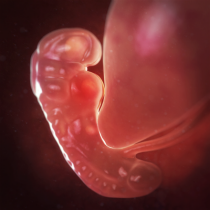
Week 11: From embryo to fetus
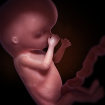
Week 11 marks an important milestone: the embryo has become a
fetus! The head is still large compared to the rest of the body. From this point on, it’ll grow more slowly than the body while continuing to become rounder. Your baby measures about 45 mm from crown to rump and weighs around 10 g.
Your little one’s face is now easy to spot. Their eyes have migrated from the sides to the front of the head and are covered by eyelids. Baby’s lips are also well defined, and they can open their mouth. They’re even drinking amniotic fluid! Fortunately, their kidneys and urinary system have begun to function. These filter the swallowed liquid and discharge it into the bladder as urine.
In the mouth and nose, the initial cellular structures have been transformed into taste buds (papillae) and smell receptors. The salivary glands and vocal cords will also begin to develop this week.
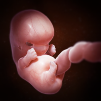
Week 12: The nails begin to grow
All of your baby’s organs and vital systems are now partially formed. Their limbs continue to become more defined as the nails start to develop. Up to this point, part of the small intestine has resided within the base of the umbilical cord. By week 12, the intestines are fully inside the abdomen.
In the brain, nerve cells are multiplying. They connect to each other by creating numerous synapses. Certain so-called primitive or archaic reflexes can already be observed. For example, your baby curls their fingers and toes if something touches their palms or the soles of their feet. This and other primitive reflexes will remain for some time before gradually disappearing. They’re an indication of proper neurological development.
Your baby now measures around 60 mm from crown to rump and weighs about 18 g.
The amniotic sac now fills your entire uterus, which is ideal for your first ultrasound. After this week, the risk of miscarriage drops considerably. If you haven’t already done so, you can start sharing the good news!
Amniotic fluid
Throughout pregnancy, your baby is surrounded by a liquid called amniotic fluid. This liquid is vital. It is contained by a thin membrane that creates a closed pocket inside your uterus. This keeps your baby’s environment at a stable temperature and allows them to move around while being protected from shocks and infections. Amniotic fluid is also a source of water and some nutrients. The most important nutrients, however, come from your blood and are delivered through the umbilical cord. When you’re ready to give birth, the amniotic sac will rupture. It contains about a litre of liquid.
Week 13: Spinal development
Your baby’s first bone tissues are forming, notably those in their pelvis, ribs, head, and limbs, though for now they’re quite soft. Their spine is also developing gradually.
At the same time, your little one’s tooth buds, soon to become baby teeth, have emerged. Their adult teeth will come in beneath these later on, during your second trimester.
Your baby’s first red blood cells are produced in the yolk sac, a round structure connected to the wall of the abdomen, and then in the liver. After a few months, their bone marrow will form.
Their delicate skin is covered with very fine little hairs known as lanugo. It usually disappears before birth. The pigment that will give their skin its colour is also developing. Thus far, your baby’s skin has been almost transparent. Finally, touch receptors now cover their entire face, their palms, and the soles of their feet.
Your baby measures about 75 mm from crown to rump and weighs around 30 g.
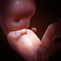
Week 14: Baby starts sucking their thumb
Your little one’s face is coming to life! Features like the nose and mouth have become more defined, and their ears are in their proper place at the sides of the head. Your baby can now make certain facial expressions like frowning and grimacing, though these are still mostly reflexes. Another distinctive feature, their fingerprints, also appear at this stage.
Your baby is now starting to react to external stimuli. If you push on your belly, they may respond by pushing back! They may also have begun sucking their thumb.
The sex of your baby will become apparent sometime this week or the next. In girls, the ovarian follicles will form, while in boys, the prostate will become visible. At your next ultrasound, you’ll be able to find out the sex of your child if you wish.
At the end of the first trimester, your baby measures about 85 mm from crown to rump and weighs around 45 g.
| Scientific review: Dr. Françoise Rypens, radiologist, CHU Sainte-Justine, and Dr. Chantal Lapierre, head of medical imagery, CHU Sainte-Justine
Research and copywriting:The Naître et grandir team
Updated: May 2021
|
Photo: iStock/kupicoo
References
Please note that hyperlinks to other websites are not updated regularly, and some may have changed since publication. It is therefore possible that a link may not be found. If a link is no longer valid, use search engines to find the relevant information.
-
Public Health Agency of Canada “The Health Pregnancy Guide.” 2011. phac-aspc.gc.ca
-
Campbell, Stuart. Échographie mode d’emploi. Éditions Marabout, France, 2005.
-
Fortier, Marie. Mes cours prénataux. Les Éditions Caractère inc., Montreal, 2014, 278 pp.
-
HANPRASERTPONG, T. et V. Phupong. First trimester embryonic/fetal heart rate in normal pregnant women. Archives of Gynecology and Obstetrics, 2006, 274, 257-260.
-
Ladewig, Patricia, et al. Maternal & Child Nursing Care. 3rd ed., Upper Saddle River, Prentice Hall, 2011, 2,016 pp.
-
Larousse. Vous et votre grossesse. Éditions Larousse, France, 2002.
-
Mayo Clinic. “Pregnancy week by week - Fetal development: The 3rd trimester.” mayoclinic.com
-
Raisingchildren.net.au. “Pregnancy: Week by week.” raisingchildren.net.au
-
Universities of Fribourg, Lausanne, and Bern (Switzerland). “Online course in embryology for medicine students.” www.embryology.ch
-
NOVA PBS. “Life’s Greatest Miracle.” pbs.org
|